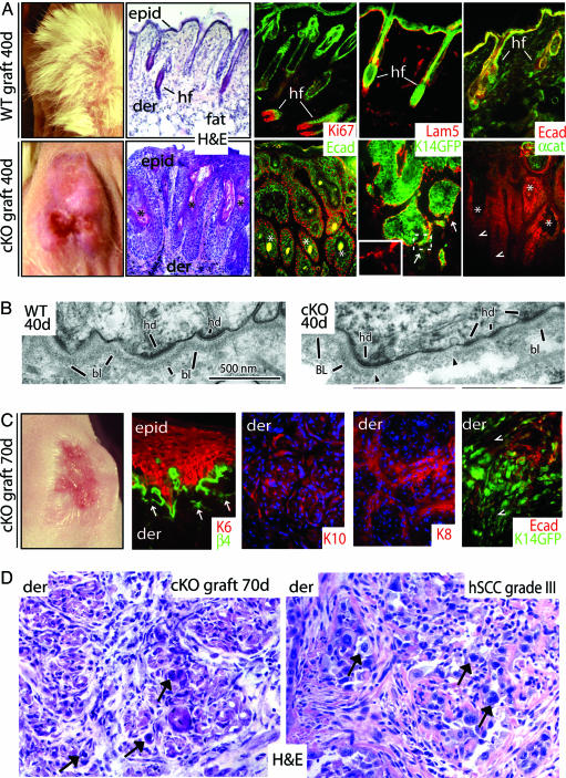
Fig. 1.
Progression to SCC in the absence of α-catenin. E18.5 WT and cKO skins were grafted onto Nude mice for the days indicated. Seventy-day WT grafts were analogous to 40 days and are not shown. (A, C, and D) Far left images in A and C are of grafts (note the absence of follicles in cKO grafts). Hematoxylin and eosin (H&E)-stained sections are from grafts, with the exception of D Lower Right, which is of a grade III hSCC (arrows denote atypical cells, classical morphological signs of SCC, and prevalence in the dermis of the 70-day cKO graft). Color coding of immunostained sections is according to secondary Abs; some sections are counterstained with DAPI (blue). Note that in C most sections are of invasive keratinocytes within the dermis as indicated in upper left of each frame. Arrows in A and C denote perturbations at dermo-epidermal borders (hemidesmosome/basement membrane), more extensive at 70 days than 40 days. Arrowheads denote epidermal cells displaying weak or no E-cadherin, which in C are also weak for K14-GFP+. epid, epidermis; der, dermis; hf, hair follicles; Lam5, laminin-5; Ecad, E-cadherin; αcat, α-catenin; K14GFP, transgenic epifluorescence expression of GFP; Ki67, a proliferating nuclear antigen; K, keratin. Asterisks denote keratinized pearls. (B) EM of ultrathin sections that correspond to boxed areas in Fig. 6 A′ and B′. Shown are the dermo-epidermal boundaries. Note that the basal lamina (bl) and hemidesmosomes (hd) are intact in WT but are perturbed in cKO (region flanked by arrowheads).

