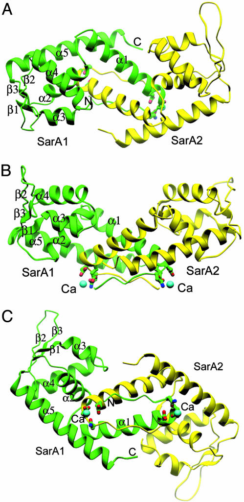
Fig. 1.
Structures of the SarA dimer, side chains of residues that involve in Ca2+ binding are shown on the convex side of the SarA dimer. The assumed DNA-binding surface is opposite of Ca2+-binding sites. Two β-hairpins (β2 and β3 from monomer one, and those from monomer two) two HTH motifs (α3 and α4 from monomer one, and those from another monomer) build up the putative DNA-binding surface on the concave side of the SarA dimer. All structural model figures are made by program ribbons (21).

