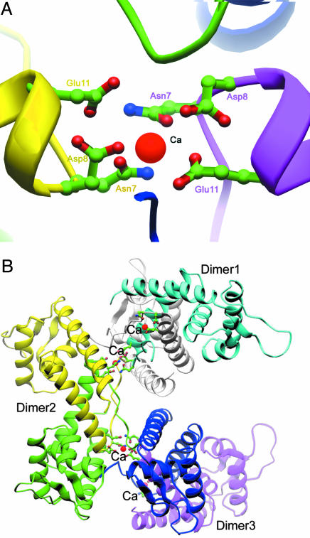
Fig. 2.
The positions of Ca2+ and their association with multiple dimers of the SarA. (A) Detailed side chains of residues that involve Ca2+ binding are shown from both dimer of SarA; calcium ions are colored purple. (B) The association of dimers of SarA through Ca2+; three SarA homodimers are involved through an x-ray axis (61).

