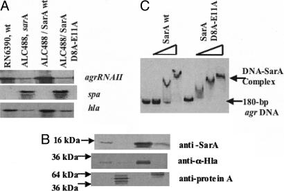
Fig. 5.
Effects of sarA mutations at codon positions 8 and 11 in in vivo and in vitro expression of target genes. (A) Northern blots of RNA isolated from S. aureus RN6390, isogenic sarA mutant ALC488, and ALC488 containing shuttle plasmid with the wild-type and mutated sarA fragments and probed with agr RNAII, spa or hla probe. (B) Western blots for intracellular, secreted spent supernatant and cell-wall associated protein extracts were probed with monoclonal anti-SarA antibody 1D1, polyclonal sheep anti-α-hemolysin antibody and affinity-purified chicken anti-protein A antibody, respectively. Equivalent amounts of extracts from the same growth conditions were used to detect SarA and protein A expression, whereas equal OD650 of spent cell culture supernatants were precipitated to detect α-hemolysin expression. Lanes are indentical to A. (C) Autoradiogram of an 8.0% nondenaturing gel showing gel-mobility shifts for increasing amounts (0, 50, 100, and 300 ng) of purified wild-type and mutated SarA proteins with a 180-bp agr P2-P3 promoter fragment (0.1 ng per reaction).

