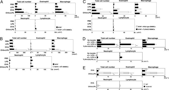
Fig. 2.
TLR4 on mast cells is required for LPS-induced enhancement of eosinophilic inflammation. (A) BMMCs (1 × 10⁷) were transferred intravenously 2 days before the initial OVA sensitization. The mean values, with standard deviation, of numbers of total cells and each cell type in the BAL fluid of W/Wv mice reconstituted with wild-type (+/+) BMMCs are shown. Ten mice were used in each group. ∗∗, <0.01 (Dunnett multiple comparisons test). (B) Histological analysis of the lung with H.E. and Luna staining. Mean values, with standard deviation, of infiltrated cells in 1 mm2 are shown. Ten lung sections per mouse from three mice in each group were examined. ∗∗, <0.01 (Dunnett multiple comparisons test). (C) Infiltrated cells in BAL fluid of W/Wv mice reconstituted with wild-type (+/+) and TLR4 KO BMMCs are shown. Ten mice were used in each group. ∗∗, <0.01 (Dunnett multiple comparisons test). (D) Infiltrated cells in BAL fluid of W/Wv mice reconstituted with wild-type (+/+) and TLR4 KO BMMCs precultured with or without LPS (10 μg/ml) for 7 days are shown. No immunization was performed. Ten mice were used in each group. ∗∗, P < 0.01 (Student t test). (E) Wild-type and TLR4 KO mice were immunized and challenged, and infiltrated cells in the BAL fluid were assessed as in Fig. 1A. ∗, P < 0.05 (Student t test).

