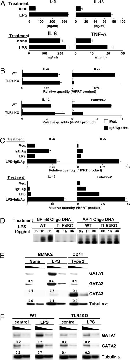
Fig. 3.
Cytokine-expression profiles and NF-κB activation in BMMCs treated with LPS. (A) BMMCs were cultured with LPS for 1 week. Then, BMMCs were restimulated with PMA (10 ng/ml) and ionomycin (1 μM) for 24 h to assess the production of IL-5, -13, and -6 and TNF-α by ELISA. Four experiments with individual BMMC preparations were performed, and similar results were obtained. (B) Wild-type (+/+) and TLR4 KO BMMCs were cultured with LPS for 1 week and then stimulated with IgE/Ag. Transcriptional levels of IL-4, -5, and -13 and Eotaxin-2 were determined by real-time RT-PCR analysis. Two independent experiments were performed, and similar results were obtained. (C) Wild-type BMMCs were stimulated with combinations of LPS and IgE/Ag. Transcriptional levels of IL-4, -5, and -13 and Eotaxin-2 were determined by real-time RT-PCR analysis. Two independent experiments were performed, and similar results were obtained. (D) EMSAs for NF-κB and AP-1. Nuclear extracts of wild-type and TLR4 KO BMMCs treated with LPS for 1 and 3 h were subjected to EMSAs with NF-κB and AP-1 probes. Two independent experiments were performed, and similar results were obtained. (E) The levels of protein expression of GATA1 and -2 in LPS-stimulated BMMCs. BMMCs treated with LPS for 3 days and CD4 T cells cultured under Th2-skewed conditions for 3 days were prepared. Nuclear extracts were used for immunoblotting with specific mAbs specific for GATA1, -2, and -3. Arbitrary densitometric units normalized with the band intensity of tubulin α are shown under each band. Three experiments were performed, and similar results were obtained. (F) GATA1 and -2 expression in LPS-treated BMMCs from wild-type and TLR4 KO mice. Two experiments were performed, and similar results were obtained.

