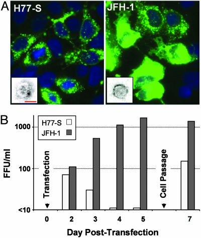
Fig. 2.
Infection of Huh-7.5 cells with H77-S and JFH-1 virus released into supernatant fluids of transfected Huh-7.5 cells. (A) HCV core antigen expression in cells infected with H77-S (Left) or JFH-1 (Right) virus. Left Inset shows a particle with immunogold labeling indicating recognition by the AP33 mAb to E2. (Bar: 50 nm.) Right Inset shows a typical JFH-1 particle for comparison. (See also Fig. 5.) (B) Time course of infectious H77-S (open bars) and JFH-1 (filled bars) virus released into supernatant fluids of RNA-transfected Huh-7.5 cells. H77-S release was greatest 24–48 h after transfection or passage of cells.

