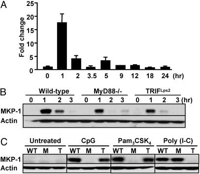
Fig. 2.
TLR stimulation induces MKP-1 expression in macrophages. (A) BMDM from WT mice were treated with 10 ng/ml LPS for the indicated time points. RNA was isolated and subjected to quantitative RT-PCR analysis of MKP-1 levels. (B) BMDM from WT, MyD88−/−, and TRIFLps2 mice were treated with 10 ng/ml LPS, and whole-cell lysates were subjected to Western blot analysis using anti-MKP-1 and anti-Actin Abs. (C) BMDM from WT, MyD88−/− (M), and TRIFLps2 (T) mice were stimulated with 1 μM CpG, 50 μg/ml poly(I-C), or 200 ng/ml Pam3CSK4 for 1 h, and MKP-1 protein expression was examined as above.

