Fig. 5.
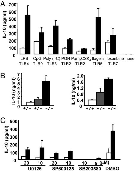
Enhanced IL-10 production in MKP-1−/− macrophages can be blocked by a p38 MAPK inhibitor. (A) BMDM from MKP-1+/+ (open bars) and MKP-1−/− (filled bars) mice were stimulated with 10 ng/ml LPS, 1 μM CpG, 50 μg/ml poly(I-C), 10 μg/ml peptidoglycan (PGN), 200 ng/ml Pam3CSK4, 100 ng/ml flagellin, or 200 μM loxoribine, or left untreated for 12 h. IL-10 production was measured by ELISA. (B Left) Serum from MKP-1+/+ (white bars), MKP-1+/− (gray bars), and MKP-1−/− (black bars) mice (n ≥ 3 for each group) was collected 3 h after LPS challenge, and IL-10 levels were measured by ELISA. (Right) BMDM from MKP-1+/+, MKP-1+/−, and MKP-1−/− mice were stimulated with 10 ng/ml LPS for 12 h, and IL-10 levels were measured by ELISA. (C) BMDM from MKP-1+/+ (open bars) and MKP-1−/− (filled bars) mice were treated with U0126 (ERK inhibitor), SP600125 (JNK inhibitor), SB203580 (p38 MAPK inhibitor), or vehicle alone (DMSO) at the indicated concentrations for 30 min and activated with 10 ng/ml LPS for 12 h. IL-10 levels were measured by ELISA.

