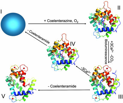
Fig. 1.
The three-dimensional structures of conformation states of obelin: II, obelin with 2-hydroperoxycoelenterazine in the absence of Ca2+ (PDB ID code 1EL4) (15); III, Ca2+-discharged obelin with both coelenteramide and bound Ca2+ (present result); IV, Ca2+-discharged obelin with coelenteramide but without Ca2+ (PDB ID code 1S36) (17); V, Ca2+-loaded apo-obelin (PDB ID code 1SL7) (18). The structure of apo-obelin (state I) is not determined. The crystal structures of different obelin conformation states are presented in the same orientation. The 2-hydroperoxycoelenterazine and coelenteramide molecules are displayed by the stick models in the center of the protein; the calcium ions are shown as red balls.

