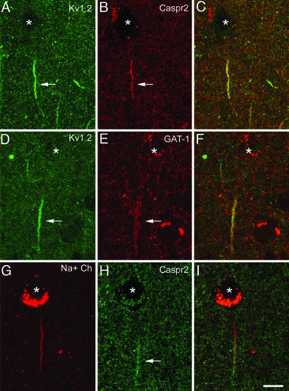
Fig. 3.
Confocal images from the same section and microscopic field illustrating double labeling for Kv1.2 and Caspr2 (A–C), Kv1.2 and GAT-1 (D–F), and VGSC and Caspr2 (G–I) in the human temporal neocortex. The asterisks indicate the location of the pyramidal cell bodies. C, F, and I were obtained after combining the images A and B, D and E, and G and H, respectively. Note that Kv1.2 immunostaining is confined to the distal region of the AIS where Caspr2 is also expressed (filled arrows in A and B). This distal AIS specifically receives GABAergic innervation from the GAT-1-ir Ch-terminals (filled arrows in D and E). VGSC and Kv1.2 partially overlap at the distal AIS (filled arrow in F), whereas the proximal AIS is characterized by the presence of VGSC and the absence of Kv1.2 (D–I). Images A–C represent stacks of nine optical sections obtained at a distance of 1.1 μm in the z axis (total: 9 μm). Images D–F and G–I represent stacks of four optical sections obtained at a distance of 1.1 μm in the z axis (total: 3 μm). (Scale bar: 11 μm.)

