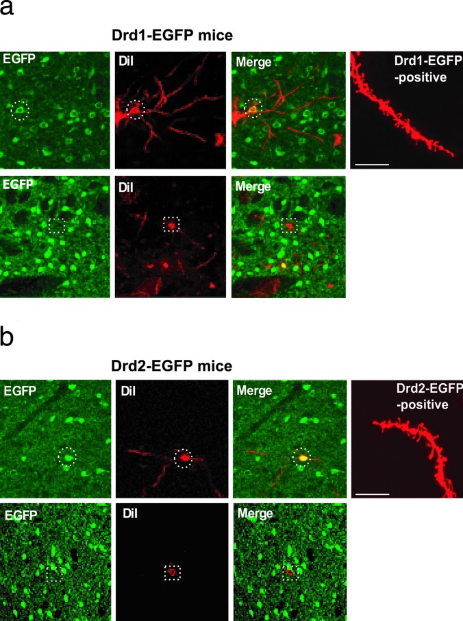
Fig. 2.
Analysis of dendritic spines in Drd1-EGFP and Drd2-EGFP mice. Neurons in NAcc of either Drd1-EGFP mice (a) or Drd2-EGFP mice (b) were first labeled with DiI (red) and then subjected to immunohistochemistry using an anti-GFP antibody (EGFP, green). Only MSNs were labeled with DiI. Images of DiI staining and GFP staining were overlaid (Merge). (a Upper) The dashed circles show an example of double labeling of a DiI-positive and Drd1-EGFP-positive cell. (a Lower) The dashed squares show a lack of double labeling of cells in slices from Drd1-EGFP mice. (b Upper) The dashed circles show an example of double labeling of a DiI-positive and Drd2-EGFP-positive cell. (b Lower) The dashed squares show a lack of double labeling of cells in slices from Drd2-EGFP mice. The rightmost images in a Upper and b Upper show examples of DiI-stained distal dendrites in Drd1-EGFP or Drd2-EGFP mice, respectively. (Scale bars: 10 μm.) The examples shown were from saline-treated mice.

