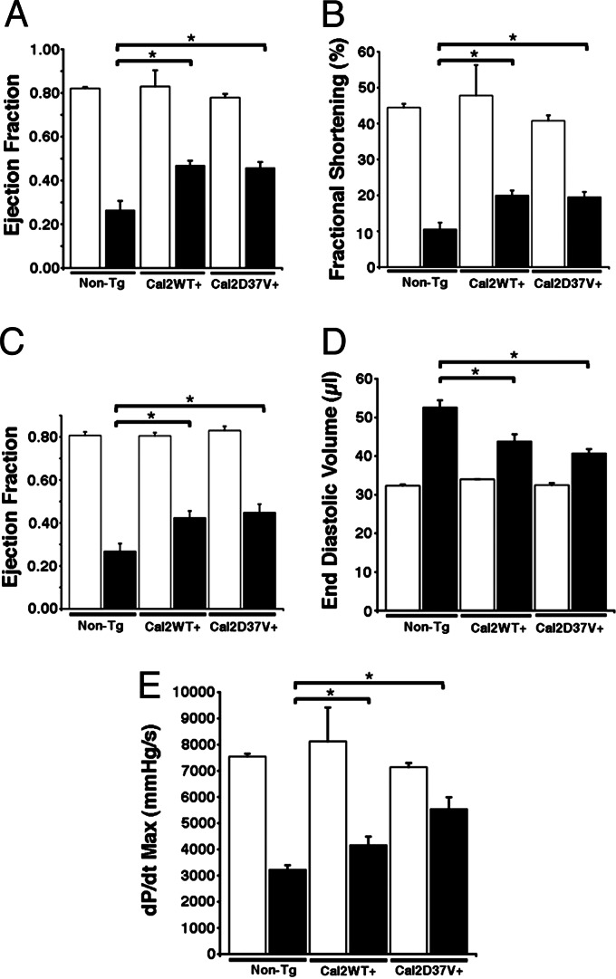
Fig. 6.
Evaluation of mouse cardiac function 6 weeks after MI. (A and B) Ejection fraction (A) and fractional shortening (B) assessed by M-mode echocardiography exhibited improved cardiac function in WT calstabin2+ and calstabin2–D37V+ mice compared with non-Tg mice after MI. (C) Hemodynamic measurements showed significant preservation of cardiac function in Tg mice compared with WT. (D) End diastolic volume was decreased in Tg mice as measured by pressure–volume loop analysis consistent with reduced dilatation after MI. (E) Maximum dP/dt was increased in Tg mice consistent with improved cardiac contractility after MI. Black bars represent infarcted animals, and white bars represent noninfarcted, sham-operated mice. ∗, P < 0.05 (significance compared with non-Tg group).

