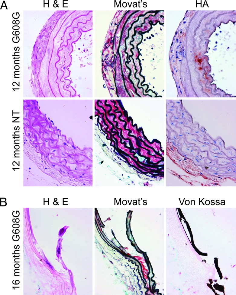
Fig. 2.
Progressive arterial abnormalities in progeria mice. (A) Analysis of aortic sections of 12-month-old mice by using hematoxylin/eosin (H & E) and Movat's pentachrome stain and immunohistochemistry analysis for hyaluronan indicates a progressive loss of VSMC, elastin breakage, thickened medial layer and adventitia, and PG accumulation. (B) All G608G mice examined over 16 months of age showed evidence of carotid (shown) and aortic (data not shown) calcification, which was confirmed with Von Kossa staining. Younger HGPS and all ages of NT mice showed no evidence of calcification. HA, hyaluronan.

