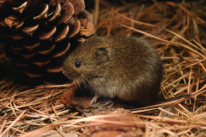
A fruitful collaboration between behavioral and molecular biologists has produced a fascinating look at the internal circadian organization of the common vole (Fig. 1). The article by van der Veen et al. (1) published in this issue of PNAS documents the relationship between circadian rhythms in the brain and those in the liver of this small rodent, whose field behavior and unusual feeding habits have been well studied but whose molecular biology has been almost completely ignored.
Fig. 1.
Meadow vole, Microtus pennsylvanicus.
Our understanding of circadian biology has undergone at least two revolutions in the last decade. The most dramatic has been the rapid increase in information and ideas about the molecular mechanisms that generate the basic near-24-h oscillations. These may have evolved three or four times in the long history of life on Earth, in bacteria, plants, perhaps fungi, and animals (2). In animals, many aspects of the mechanism have been conserved at least from flies to humans (3).
The second revolution has grown out of the demonstration, long predicted without direct evidence, that most organs, tissues, and even cells of multicellular organisms contain independent circadian oscillators that continue to oscillate when removed from the body (4–6). To a large degree this second revolution has depended on the first, because most of the assays that have made it possible to measure circadian oscillations in cells and tissues involve measuring the expression of known “clock” genes. The emerging picture of organisms as ensembles of distributed circadian oscillators has raised many important questions about how such systems are organized. Is there a strict hierarchy from a single central oscillator in the brain to peripheral structures, or, as could have been predicted and now seems clear, are there multiple central oscillators, different signaling pathways to the oscillators in the periphery, and many feedback loops? These issues are under active investigation in many laboratories around the world and promise new insights into how physiological processes are regulated.
The next revolution will require a deep understanding of the ways in which the circadian system functions adaptively in the real world. For that we need to move from the study of “virtual” animals like laboratory rats and mice to real animals. Enter the voles. Although they look superficially like mice, voles are very different creatures. In fact, metabolically they are more like cows, turning grass into meat with high efficiency, which makes them a favorite snack or a staple in the diet of many different predators. They are eaten by foxes, coyotes, wolves, raptors, cats, snakes, largemouth bass (!), and probably anything else that likes meat and can catch them. Different species of voles have evolved diverse strategies for dealing with their unenviable position in the food chain. Often these strategies involve the capacity for explosive reproduction. One particularly charming species, Microtus ochrogaster, forms lifelong, monogamous pair bonds and likes to cuddle (7, 8). Coupled with rapid fetal and neonatal development and a postpartum estrus, this bonding ensures that the females are always both nursing and pregnant!
The species studied by Schibler and colleagues (1), Microtus arvalis, has evolved an unusual antipredator strategy: the entire population, over a wide area, emerges synchronously from their burrows to feed every 2–3 h (9). The adaptive significance of this feeding pattern lies, at least in part, in “predator swamping.” The predators are confused by so many opportunities and find it hard to focus on a single animal. Gerkema and colleagues (10) have studied possible mechanisms of synchronization of these feeding cycles and have concluded that the animals have “light-insensitive ultradian (i.e., 2–3 h) oscillators … reset every dawn by the termination of the activity phase controlled by the circadian pacemaker, which is itself entrained by the light–dark cycle.” This pattern of feeding and activity raises intriguing questions about rhythmicity of central and peripheral oscillators in these animals.
We know from experiments with rats and mice that there are circadian oscillators in the liver that respond rapidly to cycles of food availability (11, 12). In fact, food intake resets the liver clock more quickly than does the light cycle, and, conversely, the light cycle resets the suprachiasmatic nucleus (SCN, the brain clock that controls most behavior) more quickly than does food (12). The data suggest that the two clocks are normally held in adaptive synchrony by SCN regulation of rhythmic feeding behavior, which in turn produces rhythmic food intake that entrains the liver clock (13). What happens to this relationship when an animal eats every 2–3 h around the clock?
Schibler and colleagues (1) housed M. arvalis singly in a 12-h light/12-h dark cycle in laboratory cages with ad lib food and recorded their locomotor activity in two different ways, with and without access to a running wheel. Animals without access to a running wheel had ultradian activity and feeding bouts every 2–3 h throughout the day and night. The availability of a wheel changed this pattern dramatically; most activity was at night and the ultradian pattern was suppressed. They then used several different molecular techniques to analyze the temporal expression of clock and clock-controlled genes in SCN and liver. In the SCN, expression was “circadian”† and appropriately phased to the light–dark cycle under all housing and feeding conditions, confirming that, in this animal as in the laboratory mouse, the light cycle was the dominant synchronizer for the SCN. In contrast, gene expression in the liver strongly depended on feeding/housing conditions. In ad lib-fed voles housed without a wheel, liver gene expression, like locomotor activity, was arrhythmic in the circadian range (the sampling intervals for gene expression measurements made it impossible to tell whether gene expression was ultradian). When circadian rhythmicity was imposed on the voles, either by giving them access to a wheel or restricting their feeding to 16 h per 24-h cycle, gene expression in the liver became weakly circadian in the first case and strongly so in the second. Similar results were obtained by measuring gene expression in the kidney.
The liver’s circadian arrhythmicity in response to the wheel-less condition, which allows the expression of ultradian feeding bouts as in the field, suggests that it is metabolically important for these animals to suppress circadian rhythmicity in the liver, although not in the brain, to deal with the digestive demands of frequent meals (cf. ref. 14, which documents suppression of circadian rhythmicity in arctic reindeer). Importantly, laboratory mice forced to feed in an ultradian pattern like that of voles retained their circadian rhythms of liver gene expression, albeit at somewhat reduced amplitudes. These findings suggest that the temporal pattern of the liver’s response to food intake is species-specific and has evolved in response to the metabolic demands of particular life styles. We do not know the extent to which such adaptations characterize other aspects of internal circadian organization, but by using old-fashioned comparative approaches coupled with modern molecular techniques, we now have the capacity to find out.
See companion article on page 3393.
Footnotes
Conflict of interest statement: No conflicts declared.
I have followed the authors’ practice of calling these rhythms circadian, although light cycles were present throughout the experiments. Although not strictly correct, other terminology would be awkward, and there is ample justification for this usage from other studies.
References
- 1.van der Veen D. R., Le Minh N., Gos P., Arneric M., Gerkema M. P., Schibler U. Proc. Natl. Acad. Sci. USA. 2006;103:3393–3398. doi: 10.1073/pnas.0507825103. [DOI] [PMC free article] [PubMed] [Google Scholar]
- 2.Bell-Pedersen D., Cassone V. M., Earnest D. J., Golden S. S., Hardin P. E., Thomas T. L., Zoran M. Nat. Rev. Drug Discovery. 2005 Jun 10; doi: 10.1038/nrg1633. 10.1038/nrd1633. [DOI] [PMC free article] [PubMed] [Google Scholar]
- 3.Young M. W., Kay S. A. Nat. Rev. Genet. 2001;2:702–715. doi: 10.1038/35088576. [DOI] [PubMed] [Google Scholar]
- 4.Balsalobre A., Damiola F., Schibler U. Cell. 1998;93:929–937. doi: 10.1016/s0092-8674(00)81199-x. [DOI] [PubMed] [Google Scholar]
- 5.Yamazaki S., Numano R., Abe M., Hida A., Takahashi R. I., Ueda M., Block G. D., Sakaki Y., Menaker M., Tei H. Science. 2000;288:682–685. doi: 10.1126/science.288.5466.682. [DOI] [PubMed] [Google Scholar]
- 6.Yoo S. H., Yamazaki S., Lowrey P. L., Shimomura K., Ko C. H., Buhr E. D., Siepka S. M., Hong H.-K., Oh W. J., Yoo O. J., et al. Proc. Natl. Acad. Sci. USA. 2004;101:5339–5346. doi: 10.1073/pnas.0308709101. [DOI] [PMC free article] [PubMed] [Google Scholar]
- 7.Carter C. S., DeVries A. C., Getz L. L. Neurosci. Biobehav. Rev. 1995;19:303–314. doi: 10.1016/0149-7634(94)00070-h. [DOI] [PubMed] [Google Scholar]
- 8.Aragona B. J., Liu Y., Yu Y. J., Curtis J. T., Detwiler J. M., Insel T. R., Wang Z. Nat. Neurosci. 2006;9:133–139. doi: 10.1038/nn1613. [DOI] [PubMed] [Google Scholar]
- 9.Gerkema M. P., van der Leest F. J. Comp. Physiol. A. 1991;168:591–597. doi: 10.1007/BF00215081. [DOI] [PubMed] [Google Scholar]
- 10.Gerkema M. P., Daan S., Wilbrink M., Hop M. W., van der Leest F. J. Biol. Rhythms. 1993;8:151–171. doi: 10.1177/074873049300800205. [DOI] [PubMed] [Google Scholar]
- 11.Schibler U., Ripperger J., Brown S. A. J. Biol. Rhythms. 2003;18:250–260. doi: 10.1177/0748730403018003007. [DOI] [PubMed] [Google Scholar]
- 12.Stokkan K. A., Yamazaki S., Tei H., Sakaki Y., Menaker M. Science. 2001;291:490–493. doi: 10.1126/science.291.5503.490. [DOI] [PubMed] [Google Scholar]
- 13.Davidson A. J., Yamazaki S., Menaker M. In: Novartis Foundation Symposium 253: Molecular Clocks and Light Signaling. Chadwick D. J., Goode J. A., editors. Chichester, U.K.: Wiley; 2003. pp. 110–125. [Google Scholar]
- 14.van Oort B. E., Tyler N. J., Gerkema M. P., Folkow L., Blix A. S., Stokkan K. A. Nature. 2005;438:1095–1096. doi: 10.1038/4381095a. [DOI] [PubMed] [Google Scholar]


