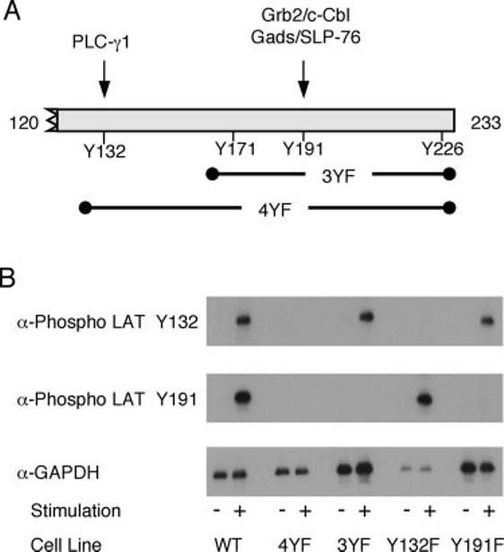
FIGURE 1.

Specificity of the anti-phospho-LAT Abs. A, Schematic of LAT showing the four C-terminal tyrosine phosphorylation sites along with the proteins that interact with LAT tyrosines 132 and 191. Also shown are the tyrosines mutated to phenylalanine in the 3YF and 4YF mutations. B, JCaM 2.5 cells transfected with various forms of LAT were treated with or without anti-CD3 for 2 min. The phosphorylation of LAT tyrosines 132 and 191 and the expression levels of GAPDH were assessed by immunoblotting as described in Materials and Methods.
