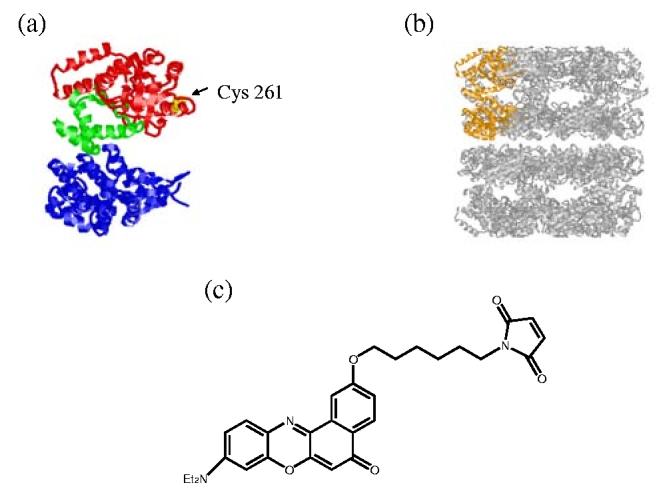
FIGURE 1.

The GroEL tetradecamer, which consists of two rings, is illustrated on the right (Figure 1B), and one of the monomer subunits shown in orange is expanded on the left to indicate the position of the mutation (Figure 1A). This cysteine mutant (Cys261, shown in yellow) was labeled with Nile Red maleimide. Color of the left figure (Figure 1A) corresponds the three different domains in GroEL; apical is red, intermediate is green and equatorial is blue. This crystal structure is from the x-ray structure of apo-GroEL, PDB filename 1GRL.6
