FIGURE 4.
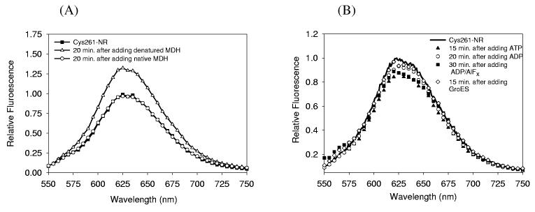
Fluorescence spectra of the Nile Red-labeled mutant GroEL (Cys261-NR, 0.2 μM) before and after the single addition of substrate, GroES, and different nucleotides (532 nm excitation). (A) Cys261-NR (rectangles) was incubated with the same amount of either unfolded (open triangles) or native MDH (open circles) at 25°C, for 20 minutes. No change in fluorescence was observed upon addition of native MDH, while unfolded MDH produced a significant increase. (B) Cys261-NR was incubated with either nucleotides (ATP (triangles), ADP (open circles), or ADP/AlFx (rectangles)) or GroES (open diamonds, 25°C, incubation times as indicated). All nucleotides and GroES induced a decrease in fluorescence compared to nucleotide-free Cys261-NR.

