
FIG. 7.
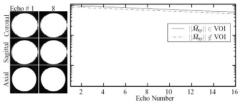
Images of a doped water phantom acquired at two of the 16 echoes of the HP-CPMG acquisition. The normalized average signal observed at each echo in representative small volumes (3 × 3 × 14 mm), one located within and one outside the VOI, is shown on a logarithmic scale.