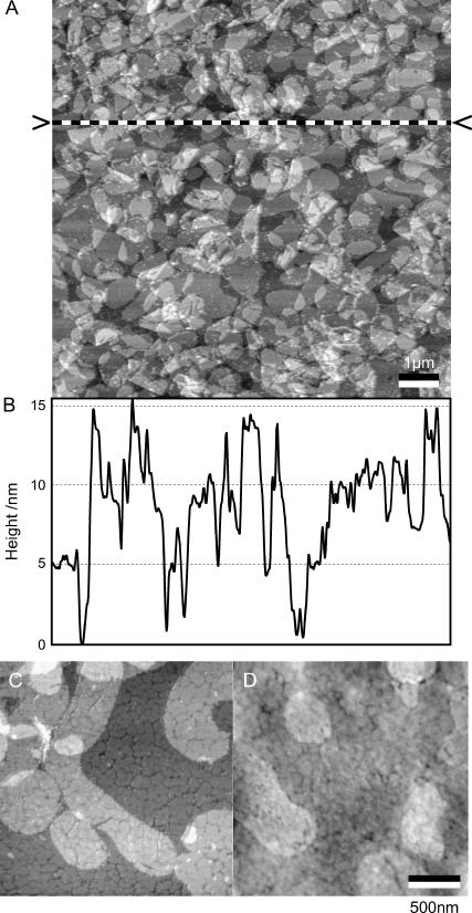
FIGURE 2.
Functionalization of the upper cantilever surface with bR membrane patches visualized by tapping mode AFM. The scale bar corresponds to 1 μm. The dashed line, also indicated by two arrowheads in panel A, corresponds to the position of the captured height profile (B). (C) Nonlabeled bR membrane patches immobilized on ultraflat gold (in air, tapping mode). (D) Immunoassayed bR patches. Antibodies are specific against the extracellular side of bR, indicating a preferential orientation of bR with the cytoplasmatic side facing the cantilever. Scale bar, 500 nm.

