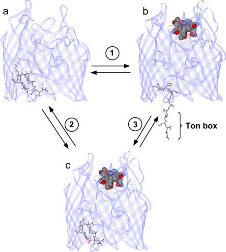
FIGURE 1.
Models for BtuB. (a) Crystal structure of BtuB in the absence of substrate (vitamin B12) (Protein Data Bank ID 1NQE), and (b) model for BtuB in the presence of substrate, where the configuration of the Ton box has been modified from that in the crystal structure to be consistent with the results of SDSL (3). In this case, SDSL indicates that the N-terminus, including the Ton box, unfolds to position 15 or 16. (c) Crystal structure of BtuB in the presence of substrate (Protein Data Bank ID 1NQH) (7). The Ton box includes residues 6–12. The portion of the structure highlighted in stick form represents residues 6–17. (1) A model for the conformational change that takes place under physiological conditions when substrate binds BtuB. (2) The conformational change that takes place under conditions of high osmolality upon the addition of substrate. (3) A conformational change that takes place upon the addition of osmolyte in the presence of substrate.

