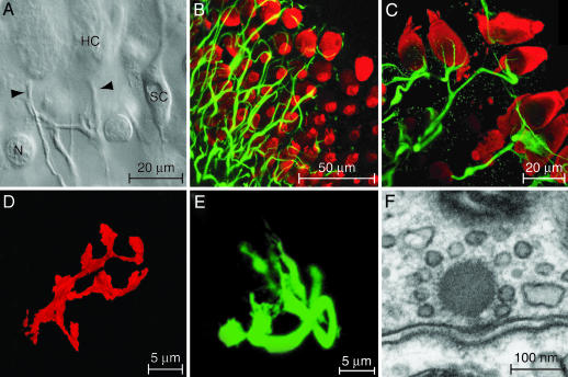
Fig. 1.
In vitro preparation of the amphibian papilla. (A) A differential-interference-contrast micrograph depicts a portion of the split sensory epithelium in which the unmyelinated eighth-nerve fibers contact the basolateral surfaces of two hair cells (HC). Arrowheads, nerve terminals; SC, supporting cell; N, nucleus of dead supporting cell. (B) A stack of confocal images of the intact epithelium depicts hair cells immunolabeled for parvalbumin 3 (red) and afferent nerve terminals labeled for neurofilament-associated proteins (green). (C) A higher-magnification view of a split preparation shows the fine branches of afferent terminals juxtaposed to the basolateral surfaces of hair cells. (D) The terminal branches of a fiber labeled with a fluorescent lipophilic tracer surround a single hair cell. (E) An afferent terminal is labeled with a fluorescent tracer after whole-cell recording. (F) As visualized by transmission electron microscopy, an afferent synapse from the amphibian papilla displays prominent pre- and postsynaptic densities and an osmiophilic presynaptic dense body, or synaptic ribbon, to which a halo of synaptic vesicles is tethered.

