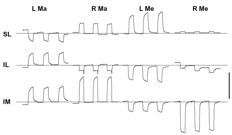
Figure 3.

Displacement of the distraction site (right side) during muscle stimulation (from #28). L, left; R, right; Ma, masseter; Me, medial pterygoid; SL, superior-lateral; IL, inferior-lateral; IM, inferior-medial. Broken lines represent the approximate baseline. Although the baseline was not always stable, the relative amount of deflection remained consistent. An upward deflection represents lengthening and a downward deflection shortening. Scale bar, 0.4 mm.
