
Figure 9. Transduction with Ad-DN-PKCɛ inhibits CCH-stimulated release of SC from lacrimal acini.
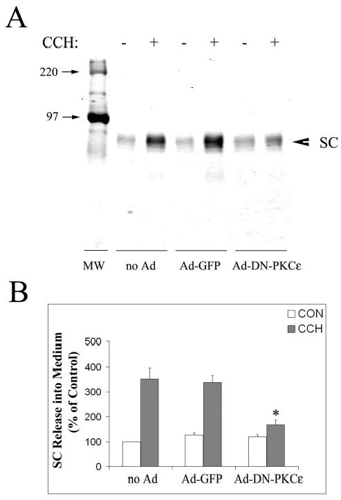
A. Western blots showing SC release into culture medium in the absence (−) and presence (+) of 100 μM CCH for 30 min in non-transduced acini (no Ad), or acini transduced with Ad-GFP or Ad-DN-PKCɛ. SC release was detected with an antibody to the extracellular, cleaved domain of pIgR combined with an appropriate IRDye™800 conjugated secondary antibody. B. SC release under each condition was quantified as shown in A., normalized to cell protein in the pellet, and compared across treatments. White bars show control (CON) release while grey bars show CCH-stimulated release. N=3 separate preparations; error bars show sem and *, significant at p ≤ 0.05 from samples co-transduced with Ad-GFP.