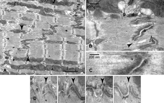
Figure 1.
(A) Electron micrograph of part of an intercalated disc between two cardiac cells showing the stairlike steps across the cells. Arrows indicate how the axial extremes of one of the transverse regions correspond to the position of the Z-discs of neighboring myofibrils. (B) Higher magnification of one transverse region showing adherens junctions (a), desmosomes (d), gap junction (g), and general apparently unspecialized plasma membrane at the tip of the folds (arrowhead). Also notable is the absence of Z-discs when myofibrils feed into the ID (open arrowhead). (C) Higher magnification view illustrating the continuity of thin filaments from sarcomere to ID membrane. (D) A series of four sections ∼100 nm apart showing the development of one of the folds (arrowhead) and the absence of plaque at the top of the fold in the final image.

