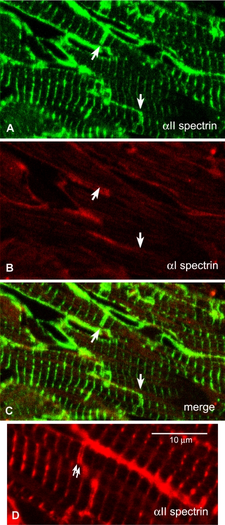
Figure 3.
(A-C) Cryosections labeled with antibodies to spectrin αI and αII. Arrows indicate IDs that can be identified in αII labeled sections but not revealed by αI. (A) αII-spectrin. (B) αI-spectrin. (C) Combined image. (D) Thin section labeled with antibody to αII-spectrin showing the doublet labeling at the ID (arrows).

