Figure 6.
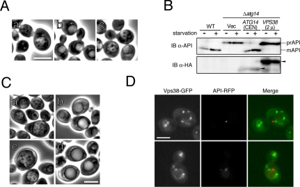
Vps38p and autophagy. (A) SEY6210 strain cells at logarithmic phase growth were transferred to S (–NC) medium in the presence of 1 mM PMSF. After 4 h of culture in S (–NC) medium, WT cells (a), Δatg14 cells (b), and Δvps38 cells (c) were observed by phase-contrast microscopy. (B) API maturation was monitored in Δatg14 cells overexpressing Vps38-HA-GFP. Cells, grown in SD + CA + AdeTrp medium at 30°C, were collected during logarithmic growth (starvation –) or 4 h after transfer to SD (–N) medium (starvation +). Lysates were subjected to immunoblotting with anti-API and anti-HA (HA-7) antibodies. (C) The accumulation of autophagic bodies was examined in Vps38-HA-GFP-overexpressing Δatg14 cells, on the BJ2168 background which lacks vacuolar proteases. WT cells (a), Δatg14 cells (b), Δatg14 cells expressing Atg14-FL from a single-copy plasmid (c), and Δatg14 cells expressing Vps38-HA-GFP from a multicopy plasmid (d) were transferred to S (–NC) medium. After 4 h of culture in S (–NC) medium, cells were observed by phase-contrast microscopy. (D) We investigated the localization of Vps38p. Diploid cells expressing chromosomal Vps38-GFP and API-RFP (YOK89) were cultured in SD + CA + AdeTrpUra medium and subjected to fluorescence microscopy. Two representative fields are shown. Bars, 5 μm.

