Figure 8.
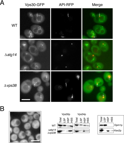
Localization of Vps30p. (A) Diploid cells expressing chromosomal Vps30-GFP and API-RFP were observed by fluorescence microscopy. The arrows indicate the structures to which Vps30-GFP and API-RFP colocalize. (B) Haploid cells expressing chromosomal Vps30-GFP (left). Vps30-GFP was dispersed throughout the cytoplasm, which was confirmed by cell fractionation analysis of WT and Δatg14Δvps38 cells expressing an untagged Vps30p (right). Dpm1p and Kex2p were used as markers for the ER and late Golgi, respectively. Bars, 5 μm.

