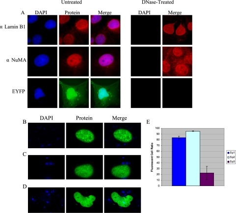
Figure 5.
Detection of nuclear matrix-associated proteins in situ using direct and indirect fluorescence microscopy. (A) Endogenous lamins (αLamin B1; H-90) and NuMA (αNuMA; N-20) were detected in untreated COS-1 nuclei as well as DNase-treated nuclear matrices via indirect immunofluorescence using Alexa Fluor 594 goat anti-rabbit and Alexa Fluor 594 rabbit anti-goat secondary antibodies, respectively. EYFP was detected in untreated nuclei via direct fluorescence. Nuclei were localized via DNA-staining (DAPI), and superimposed images are also shown (Merge). (B–D) COS-1 cells were transfected with (B) pEYFP-Sp2, (C) pEYFP-Sp1, or (D) pEGFP-Sp3, nuclear matrices were prepared with DNase I, and labeled nuclei were identified in situ. (E) COS-1 cells were transfected with expression vectors encoding pEYFP-Sp1, pEYFP-Sp2, or pEGFP-Sp3, and the total numbers of fluorescent nuclei were enumerated in untreated cultures and in nuclear matrices prepared from cultures treated with DNase I. The graph represents the ratio of total fluorescent cells recovered in DNase I–treated cultures relative to the total number detected in untreated, transfected cultures. Each transfection experiment was repeated in triplicate to account for plate-to-plate variations in transfection efficiency. Error bars, SDs.

