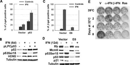
Figure 4.
Role of p53 in β-interferon–induced senescence. (A) SA-β-Gal: IMR90 cells infected with the retroviral vector pLPC or its derivative expressing p53 were treated with β-interferon or vehicle. Data were analyzed with the Student's t test. (B) Immunoblots from cells as above collected 6 d after retroviral infection. (C) SA-β-Gal: IMR90 cells were infected with the retroviral vector LXSN or its derivative expressing E6. Then the cells were treated with β-interferon or vehicle for 12 d and fixed for SA-β-Gal staining. (D) Immunoblots for cell cycle regulators and the p53 pathway in E6-expressing cells and control after treatment with β-interferon or vehicle for 12 d. (E) Colony formation assay at 39°C of TSP cells previously treated with α- or β-interferon for 8 d at 39°C or for 2, 4, or 8 d at 32°C. TSP cells expressing oncogenic ras were used as a positive control for induction of a permanent cell cycle arrest after activation of p53 at 32°C. Colonies were counted in three independent experiments, and the data were analyzed for significance with the Student's t test.

