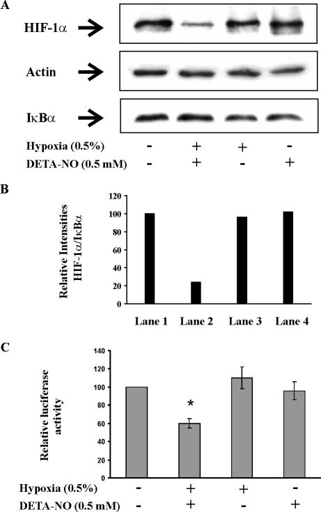
Figure 1.
Hypoxia and NO attenuate HIF-1 in RCC4 cells. (A) For protein analysis, RCC4 cells were exposed to hypoxia (0.5%), treated with 0.5 mM DETA-NO, a combination of hypoxia/DETA-NO for 4 h, or remained as controls. Western blotting was used to follow expression of HIF-1α, actin, and IκBα. Results are representative for three individual experiments. (B) For quantification of signals from A, densitrometric quantum levels of lanes 1–4 (from left to right) were measured, and relative intensities were calculated from ratios of HIF/IκBα by setting controls as 100. (C) For transactivation analysis, 2 × 10⁵ RCC4 cells were transfected with 0.5 μg of the plasmid pGLEPOHRE and exposed to hypoxia (0.5%), 0.5 mM DETA-NO, combinations thereof for 16 h or remained as controls. After cell lysis, luciferase activity was measured and normalized compared with controls. Data are the mean ± SEM (n = 3). Significant alterations are expressed relative to controls.

