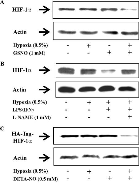
Figure 2.
Hypoxia/NO attenuates endogenous and exogenous HIF-1α expression in RCC4 cells. (A) Cells were exposed for 4 h to hypoxia (0.5%), treated with 1 mM GSNO, a combination of hypoxia/GSNO, or remained as controls. (B) Cells were exposed to hypoxia (0.5%) for 16 h, a combination of hypoxia/LPS (1 μg/ml)/IFNγ (100 U/ml) for 16 h, a combination of hypoxia/LPS/IFNγ in the presence of 1 mM l-NAME for 16 h, or remained as controls. (C) Cells were transfected with 3 μg/dish pHA-HIF-1α plasmid. Twenty-four hours later, cells were exposed for 4 h to hypoxia (0.5%), treated with 0.5 mM DETA-NO, a combination of hypoxia/DETA-NO, or remained as controls. Western blotting was used to follow expression of HIF-1α by using anti-HIF-1α or anti-HA-Tag monoclonal antibodies relative to actin. Results are representative for three individual experiments.

