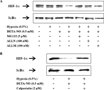
Figure 6.
Calpain mediated HIF-1α destruction. (A) RCC4 cells were exposed for 4 h to hypoxia (0.5%), 0.5 mM DETA-NO, hypoxia/DETA-NO, the combination of hypoxia/DETA-NO with the addition MG132, ALLN, ALLM, or remained as a control. (B) RCC4 cells were exposed for 4 h to the combination of hypoxia/DETA-NO, the combination of hypoxia/DETA-NO with the addition of the calpain inhibitor calpastatin peptide (2 μM), or remained as a control. Expression of HIF-1α and IκBα were followed by Western analysis. Results are representative for three individual experiments.

