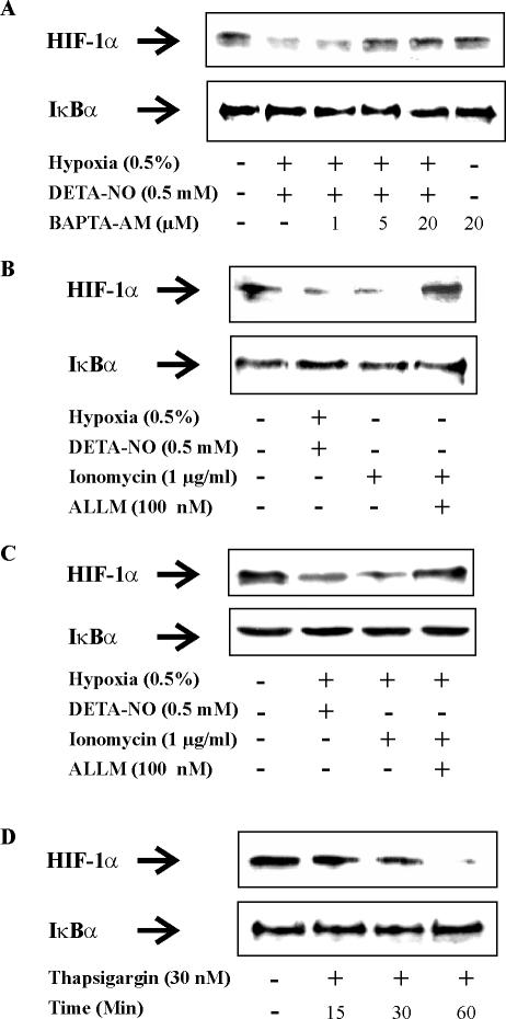
Figure 7.
Ca2+ regulates the expression of HIF-1α in RCC4 cells. (A) RCC4 cells were exposed to combinations of hypoxia (0.5%)/DETA-NO (0.5 mM) either alone or in the presence of increasing concentrations of the Ca2+ chelator BAPTA-AM for 4 h. Alternatively, cells were treated with 20 μM BAPTA-AM under normoxia, or remained as a control. (B and C) RCC4 cells were exposed for 4 h to combinations of hypoxia (0.5%)/DETA-NO (0.5 mM), or treated with 1 μg/ml ionomycin with or without the addition of the calpain inhibitor ALLM (100 nM) under (B) normoxia or (C) hypoxia, or remained as controls. (D) RCC4 cells were exposed to 30 nM thapsigargin for 15, 30, or 60 min under normoxia or left untreated. Expression of HIF-1α and IκBα were followed by Western analysis. Results are representative for three individual experiments.

