Figure 1.
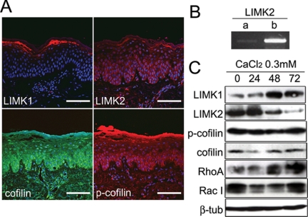
LIM kinase and cofilin expression in normal epidermis and cultured keratinocytes. (A) Normal human epidermis immunostained for LIMK1, LIMK2, cofilin, and phosphorylated cofilin with DAPI nuclear counterstain. Scale bars, 100 μm. (B) RT-PCR of primary human keratinocytes with primers specific for LIMK2a and LIMK2b. (C) Western blots of cultured human keratinocytes grown in low calcium medium (0) or transferred to medium containing 0.3 mM calcium ions for the number of hours shown. β-Tubulin was used as a loading control.

