Figure 3.
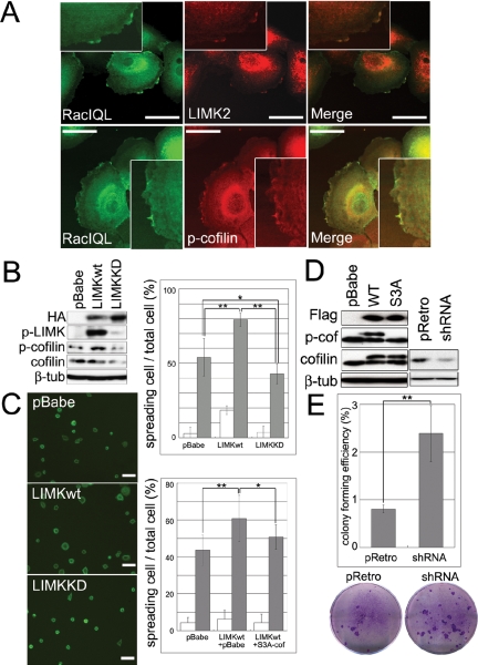
LIMK and cofilin regulate keratinocyte spreading and clonal growth. (A) Keratinocytes were infected with eGFP-Rac1 (Rac1QL), and the subcellular localizations of Rac1, LIMK2 and p-cofilin were compared by double-label immunofluorescence. Scale bars, 50 μm. Insets show cell periphery at higher magnification. (B) Western blotting of keratinocytes infected with control retroviral vector (pBabe), HA-tagged wild-type LIMK (LIMKwt), or HA-tagged kinase dead LIMK (LIMKKD). (C) Keratinocytes were plated on type I collagen for 10 min (white bars; Alexa 488-phalloidin labeling; scale bars, 100 μm) or 30 min (gray bars). Top graph shows ratio of spread cells to total adherent cells infected with pBabe, LIMKwt, and LIMKKD. Bottom graph shows ratio of spread cells to total adherent cells infected with pBabe alone, LIMKwt and pBabe, or LIMKwt and S3A-cofilin. More than 10 microscopic fields were analyzed per data set. Mean ± SEM is shown. * p < 0.05, ** p < 0.01. (D) Western blots of primary keratinocytes infected with control vector (pBabe), Flag-tagged wild-type (WT) or constitutively active (S3A) cofilin, shRNA control vector (pRetro) or shRNA for cofilin (shRNA) Upper and lower bands detected with anti-cofilin antibody are Flag-tagged and endogenous cofilin, respectively. (E) Effect of cofilin RNAi on colony forming efficiency of primary human keratinocytes. Representative culture dishes are shown. Data are mean ± SEM (** p < 0.01).

