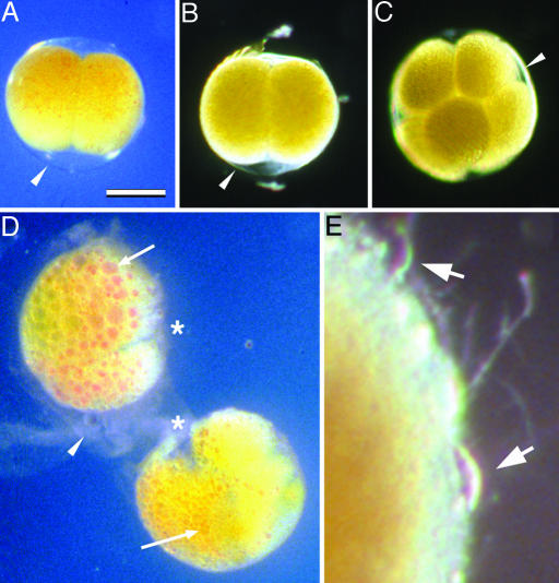
Fig. 2.
Cleavage-stage H. erythrogramma embryos under preserving and nonpreserving conditions. (A and B) Embryos killed at the two-cell stage by placing them in seawater containing 1% ammonium for 10 min, then transferring them to seawater containing 100 mM β-ME. They were photographed after 2 (A) or 10 (B) days. (C) Embryo killed at the eight-cell stage by transfer into seawater containing 100 mM β-ME, photographed after 12 days. (D) Embryos from the same two-cell stage culture as in A and B but returned to normal seawater after killing, photographed after 2 days. Embryos have undergone autolysis: cytoplasmic lipid and pigment have coalesced (arrows); cleavage furrows have degraded (asterisks); and fertilization envelopes are disintegrating (arrowhead). Autolysis is further advanced in the top embryo than in the bottom embryo. In the bottom embryo, the process is further advanced in the left-hand blastomere (arrow) than in the right-hand blastomere. (E) Decaying surface of an embryo from the set shown in A and B, returned to normal seawater after 4 days in reducing conditions, photographed 7 days later (total 11 days postdeath). Onset and progress of decay is slower than autolysis in embryos never exposed to reducing conditions. The fertilization envelope degrades and the cytoplasm of the embryo is then exposed to external decay processes, including attack by protists (arrows). (Scale bar: 200 μm for A–D; 32 μm for E.)

