
Figure 4.
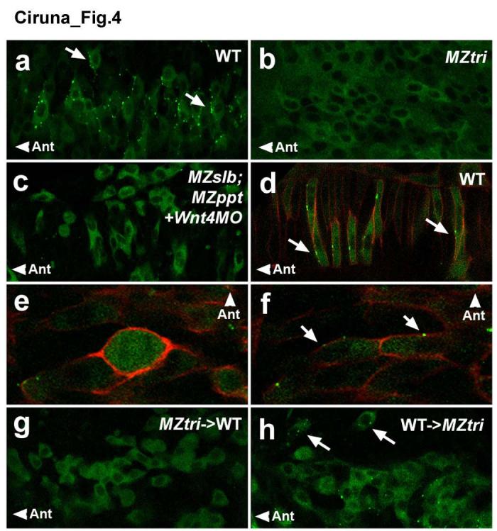
Anterior membrane localization of Gfp-Pk as a marker of planar polarity. Confocal images taken at the level of the anterior spinal cord, through the dorsal-ventral plane of the neural keel (a-c, e-h) or notochord (d) of 8- to 10-somite staged embryos. (a-c) Scatter labelling of Gfp-Pk in the neural keel of (a) a WT embryo, (b) an MZtri mutant, or (c) an MZslb;MZppt mutant injected with 6 ng of Wnt4 MO. (d) Scatter labelling of Gfp-Pk plus mRFP in WT notochord, demonstrating anterior membrane translocation of Gfp-Pk (arrows) in cells that undergo well-characterized CE movements in response to PCP signalling. (e-f) A Gfp-Pk plus mRFP labelled WT neural keel cell demonstrating transient loss of polarity markers during mitosis (e), but re-establishment of membrane-localized Gfp-Pk in both daughter cells (arrows in f). (g-h) Chimeric analysis of the autonomy of PCP signalling, using Gfp-Pk as a marker of planar polarity. (g) MZtri cells transplanted into WT host embryos do not localize Gfp-Pk to the membrane, as Vangl2 is required for Pk translocation. (h) WT cells transplanted into MZtri hosts show reduced membrane Gfp-Pk localization and abnormal polarity (arrows). “Ant” marks the anterior direction.