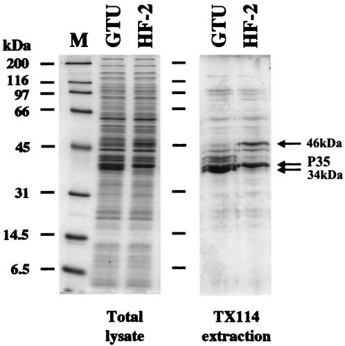
FIG. 2.
Analysis of LAMP profiles of M. penetrans. The total cell lysate and TX-114 phase-fractionated proteins of M. penetrans strains GTU and HF-2 were analyzed by SDS-12% PAGE. The proteins were stained with Coomassie blue. The positions of the P35 and 34- and 46-kDa proteins are indicated. A protein molecular mass marker is in lane M, and molecular masses are on the left.

