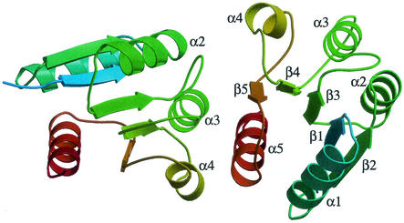
FIG. 1.
Asymmetric interface between PhoPN protomers. Each protomer is represented by ribbons, and the color varies from light blue (N terminus) to red (C terminus). The two domains associate through surface A (α4, β5, loop β5-α5, and α5) from protomer 1 (right) and surface B (α3, loop β4-α4, and α4) from protomer 2 (left) (the protomers are arbitrarily numbered). This setting leaves surfaces B and A from protomers 1 and 2, respectively, free for association with other tandem units.

