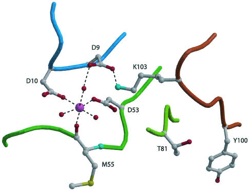
FIG. 3.
Octahedral coordination of the manganese ion in the active site of PhoPN. The Mn2+ ion is represented by a large magenta sphere. Small red spheres and dashed lines indicate water molecules and polar interactions, respectively. Outside the active site, two conserved residues in receiver domains, T81 at the C-terminal edge of strand β4 and Y100 from strand β5, are also shown.

