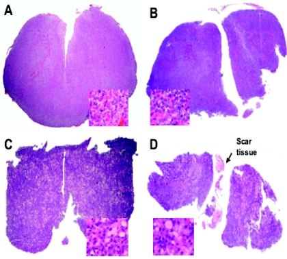
FIG. 2.
Slides of adenohypophysial tissue from sham-operated (A), NIL (B), TX (C), and NIL plus TX (D) rats with staining by the hematoxylin-eosin method (×40). No signs of infarction or other histological lesions were found in NIL rats with or without TX, except scar tissue and small isolated remnants from the intermediate lobe. The inset square in each panel shows an amplified view of the pituitary tissues (×1,000). No histological differences between sham-operated (A) and NIL (B) animals were observed. Slides of tissues from NIL rats with or without TX (C and D) showed the cells typical of thyroidectomy.

