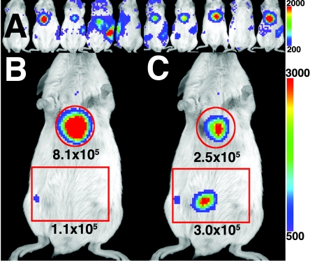
FIG. 2.
BLI of L. monocytogenes in mice after fasting and feeding. Ten CD1 mice were infected intravenously with 4 × 10⁴ CFU of the virulent 2C strain of L. monocytogenes. On day 4 postinfection, the mice were starved for 4 h and imaged (A), and the eighth mouse from the left was selected from the cohort for the fast-and-feed procedure. This animal was allowed to recover from the anesthetic for 10 min, fed 200 μl of whole cow's milk, and imaged 5 min (B) and 50 min (C) after feeding. Color bars indicate photon counts per pixel registered by the camera during the 5-min exposures, and the counts registered in the designated regions of interest (within the 1.2-cm2 red circle and the 3- by 2-cm red square) are shown. The same reference photograph was used for both BLI images.

