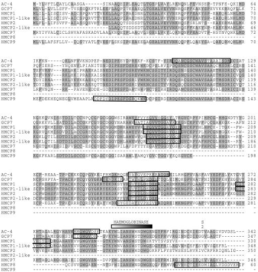
FIG. 3.
Alignment of the CBL sequences enclosing the identifications of CBLs made from ES of H. contortus. GenBank accession numbers of the displayed sequences have been indicated in Table 1. Positions having four or more identical residues have been shaded. The peptide sequences obtained from each spot by LC/MS/MS (as described in reference 28) are boxed in the alignment. The hemoglobinase motif and S2 substrate binding site described in the text are shown, and the propeptide region is indicated by a black bar.

