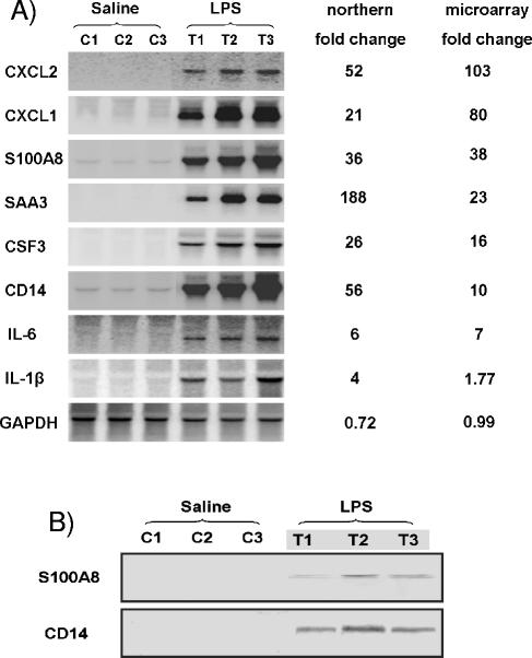
FIG. 1.
Microarray analysis of selected mammary gland gene expression changes 4 h after intramammary infusion of saline or LPS was verified by Northern blot and Western blot analyses. (A) Northern blot analysis. A total of nine representative genes were selected for this assay, as indicated. Samples of the same RNA used for microarray analysis were separated by agarose gel electrophoresis, and the resulting blots were hybridized with appropriate mouse probes. Changes (n-fold) in gene expression detected by Northern analysis were calculated by dividing the average value of LPS treatment groups (T1 to T3) by that of saline control groups (C1 to C3). The expression level of mouse GAPDH was analyzed as an internal control. (B) Western blot analysis for CD14 and S100A8. Protein (80 μg) extracted from saline-control- and LPS-treated mammary tissues (4 h post-LPS infusion) were fractionated on an SDS-12% polyacrylamide gel and then transferred to the nitrocellulose membranes. Anti-CD14 and anti-S100A8 antibodies were used.

