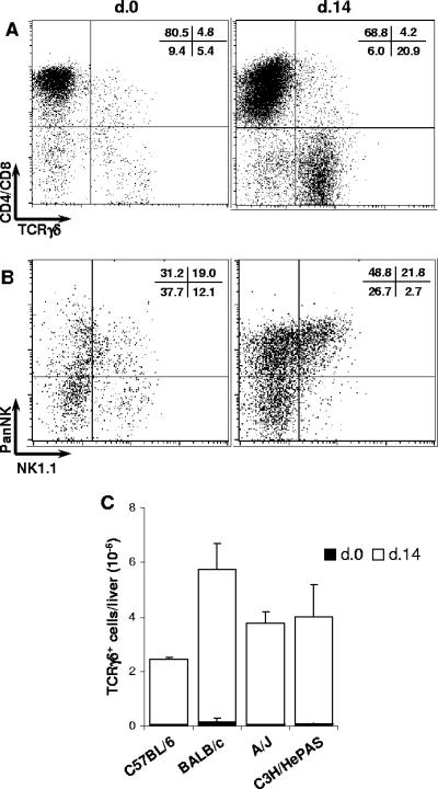
FIG. 7.
Characterization of CD4− CD8− γδ T cells in the livers of acutely T. cruzi-infected C57BL/6 mice. Fourteen days after infection, intrahepatic lymphocytes were isolated from perfused livers and analyzed by fluorescence-activated cell sorting. Noninfected animals were used as controls. In panel A, dot plots of a representative C57BL/6 mouse show the coexpression of CD4/CD8 and TCRγδ among gated CD3+ cells. Mean percentages (n = 3) of cells in each region from the same experiment (out of three experiments) are shown. In panel B, dot plots of a representative C57BL/6 mouse show the coexpression of PanNK and NK1.1 among gated TCRγδ+ cells. Mean percentages (n = 3) of cells in each region from the same experiment (out of three experiments) are shown. In panel C, total numbers of TCRγδ+ cells in the livers of mice of the four strains. Bars represent the mean ± SD (n = 3) of a representative experiment (out of two experiments).

