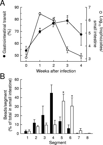
FIG. 1.
G. muris infection of wild-type mice induces small-intestinal hypermotility. (A) Adult C57BL/6 mice were infected orally with 104 G. muris cysts (weeks 1 to 4) or left uninfected as controls (week 0). At the indicated times after infection, small-intestinal motility (•) and trophozoite load (○) were examined. Motility data are shown as the distance traveled by a carmine dye-containing test meal relative to the length of the entire small intestine over a 20-min period. Values are means ± standard errors of the means (SEM) of results for four to seven animals. Asterisks represent a significant (P < 0.05, t test) increase in motility relative to uninfected controls. (B) Mice infected for 2 weeks with G. muris (empty bars) and uninfected controls (filled bars) were given a test meal containing 10-μm fluorescent polystyrene beads, and bead transit was assessed after 20 min. Bead numbers in each of eight equally sized small-intestinal segments (which are numbered in order from proximal duodenum to distalileum) are given as percentages of the total number of beads in the small intestine. No significant difference was found between the numbers of residual beads in the stomachs of uninfected and infected mice (14.2% ± 5.7% versus 18.5% ± 3.4%, respectively). Values are means ± SEM for six to seven animals. Asterisks represent a significant (P < 0.05, t test) increase relative to uninfected mice.

