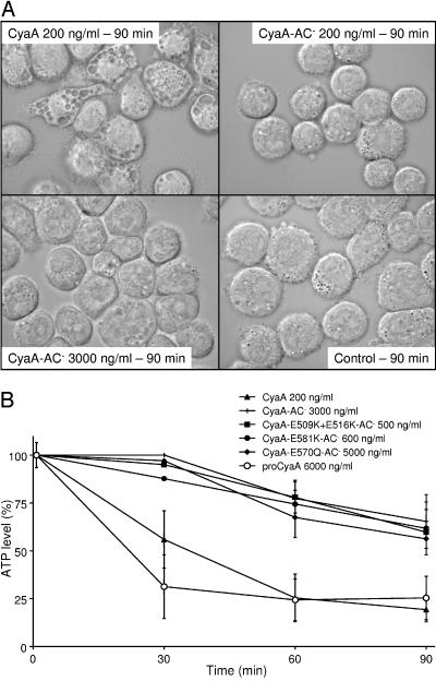
FIG. 4.
Enzymatic activity and not the pore-forming capacity of CyaA induces depletion of ATP and vacuolization of J774A.1 cells. (A) Monocytes were grown overnight on glass coverslips in RPMI medium; the medium was changed for DMEM 2 h prior to addition of the toxins at indicated concentrations. After 90 min of exposure to toxin in DMEM at 37°C, the J774A.1 cells were viewed at a magnification of ×100 using Nomarski differential interference contrast optics with an Olympus BX60 microscope. The experiment was repeated twice, and representative images from a series of micrographs are shown. (B) Pore-forming activity of CyaA causes only moderate ATP depletion even at LC50 doses of AC− toxoids. A total of 105 J774A.1 cells were incubated with mutant CyaA-AC− toxoids, intact acylated CyaA, or the nonacylated proCyaA at protein concentrations representing the respective LC50 (see Table 1) in DMEM. The ATP level in toxin-treated cells over time was determined in cell aliquots using the ATP Bioluminescence Assay kit CLS II (Roche). The results are representative of at least three independent determinations performed in triplicate.

