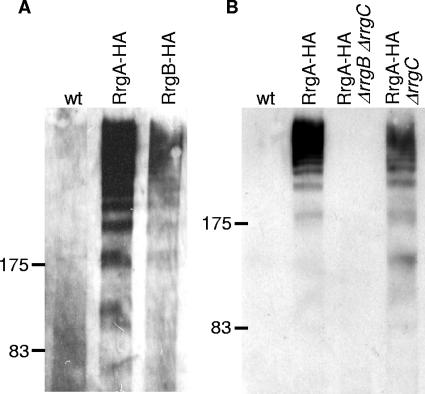
FIG. 1.
RrgA and RrgB are polymerized into pili. (A) Cell wall fractions of RrgA-HA and RrgB-HA S. pneumoniae TIGR4 form high-molecular-weight complexes when immunoblotted with anti-HA antisera. Wild-type (wt) TIGR4 (lane 1), RrgA-HA (lane 2), and RrgB-HA (lane 3) are shown. (B) The high-molecular-weight RrgA-HA complexes are not present in a strain with rrgB deleted and rrgC but are present when only rrgC is deleted. Wild-type TIGR4 (lane 1), RrgA-HA (lane 2), RrgA-HA ΔrrgB ΔrrgC (lane 3), and RrgA-HA ΔrrgC (lane 4) strains are shown.

