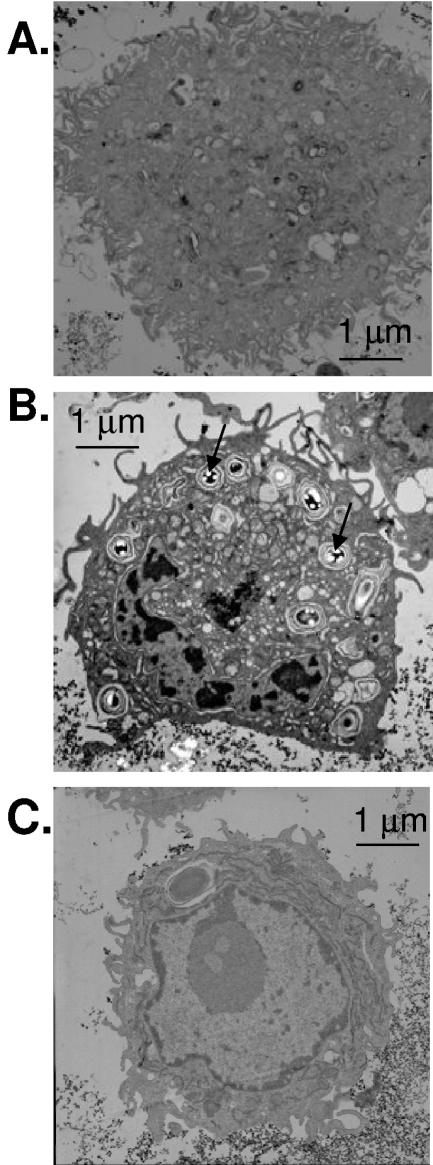
FIG. 1.
B. anthracis induces phagocytosis in RAW264.7 macrophage cells. RAW264.7 cells were infected with the ΔgerH strain of B. anthracis (MOI, 1:1) and then washed and fixed for TEM. (A) TEM of control RAW264.7 cells without infection. (B) TEM of RAW264.7 cells infected with B. anthracis ΔgerH spores for 18 h. Arrows indicate spores (light intense images) inside of phagolysosomes. (C) TEM of RAW264.7 cells pretreated for 10 min with cytochalasin D (10 μg/ml) to inhibit phagocytosis, infected with B. anthracis ΔgerH spores for 18 h, and then washed. No spores were visualized in the presence of cytochalasin D; the vacuole present in the cell is of unknown origin, but size and absence of light fragmentation suggest it is not a B. anthracis spore. The general morphology of cell organelles was not altered by exposure to B. anthracis. Bars, 1 μm.

